Skeletal Anatomy Posters
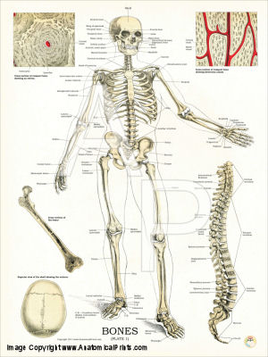
Bones - Plate 1. 18" X 24"
Human skeletal system with lateral view of the spinal column.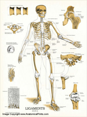
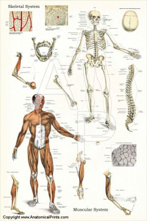
Skeleton and Muscles Poster 24" X 36"
Skeletal and Muscular Systems of the body.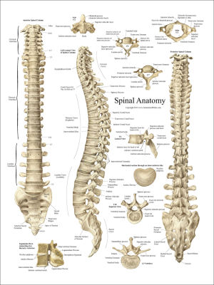
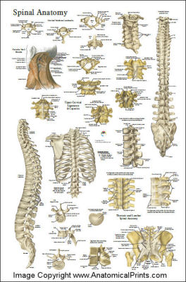
Anatomy of the Spine Poster
Spine anatomy poster shows 3 views of the spine, cervical, thoracic and luimbar vertebrae with cross-section of vertebral unit.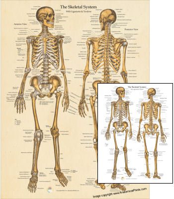
Skeletal System Poster
Anterior and posterior views of the human skeleton including ligaments and tendons.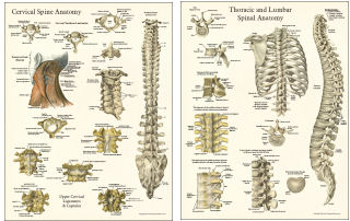
Spinal Anatomy Poster Set
Cervical and Thoracic vertebrae,; Posterior and lateral spinal column view, Ligaments of the cervical and thoracic vertebrae, Classic images from the early 1900's